Molecular Imaging
Multiplexed PET Imaging
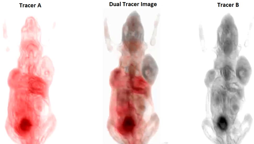
The value of positron emission tomography (PET) lies in its unsurpassed high-sensitivity for tracking biomarkers and molecular processes in vivo; however, it lacks the ability of multiplexing signals from more than one radiotracer thus limiting the amount of information available after each scan. We have developed a new technology compatible with any existing PET system, that enables simultaneous imaging of two radio tracers in a single PET acquisition. We accomplished this by combining a radiotracer labeled with a standard positron-emitting radionuclide with another labeled with a radionuclide emitting prompt γ-rays together with the positrons. Recovered images from simultaneous dual-tracer PET acquisitions were comparable to those obtained in sequential single tracer scans, allowing the identification and quantification in-vivo of several interlinked disease markers in a single acquisition
Related work
- Lage, Joaquin L. Herraiz, V. Parot. “Multiplexable Emission tomography” PCT/US2013/038846,
- Garrido, E. Lage, M. Papisov, A. Santos, M.A. Lessa, S Weise, C. Kosour, J. Venegas, J.L. Herraiz. Multiplexed dynamic PET imaging of the lungs based on triple coincidences, 2017 ISBI Conference (Asutralia)
- L. Herraiz, F. Mulero, V. Parot, S. Dave, S. Moore, J.M. Udias, J.J. Vaquero and E. Lage. “mPET: Herramienta innovadora para imagen PET multitrazador”. 34 Congreso de la Sociedad Española de Medicina Nuclear e Imagen Molecular 2014 Oral Presentation Rev Esp Med Nucl Imagen Mol. 2014;33(Supl 1):119 5-Y IF: 0.89
- L Herraiz, E. Lage, J. Venegas. “Simultaneous PET imaging of Liquid Abpsortion and Mucociliary transport in the Lungs based on Triple coincidences”. at the IEEE Nuclear Science Symposium and Medical Imaging Conference (2016 NSS/MIC), France, 2016 , Oral Presentation (#M07-4).
- L. Herraiz, S. C. Moore, M.-A. Park, J. M. Udias, J. J. Vaquero and E. Lage. “Image Quality Assessment of Multiplexed PET” IEEE Nuclear Science Symposium & Medical Imaging Conference, 2015. Oral presentation (M5C2-4).
- L. Herraiz, S. C. Moore, M. Cervo, S. Espana, J. Ruiz-Cabello and E. Lage. “Multiplexed PET in Clinical Scanners based on Triple coincidences”. IEEE Nuclear Science Symposium & Medical Imaging Conference, 2014. Oral presentation(M06-7).
- Parot, J.L. Herraiz, S. R. Dave, J.M. Udias, S.C. Moore, M. Park, J.J. Vaquero, E. Lage. “A new approach for multiplexed PET imaging”. IEEE Nuclear Science Symposium & Medical Imaging Conference, 2013. Oral Presentation (M03-5).
- L. Herraiz, E. Lage, V. parot, S. R. Dave, J.M. Udias, J.J. Vaquero, L. M. Fraile. “Production of positron-gamma emitters for multiplexed PET (mPET) imaging”. IEEE Nuclear Science Symposium & Medical Imaging Conference, 2013. Poster Presentation.
Triple coincidence imaging in PET

Triple coincidences in positron emission tomography (PET) are events in which three γ-rays are detected simultaneously. These events, though potentially useful for enhancing the sensitivity of PET scanners, are discarded or processed without special consideration in current systems, because there is not a clear criterion for assigning them to a unique line-of-response (LOR). We have developed several novel strategies to use these type of events in PET including multiplexed PET imaging, enhance the sensitivity of the scanner or improve image quality when using non-standard positron emitting radionuclides.
Related work
- E. Lage, V. Parot, S. C. Moore, A. Sitek, J. M. Udias, S. R. Dave, M. Park, J.J. Vaquero, J. L. Herraiz. “Recovery and Normalization of triple coincidences in PET”. Medical Physics 42, 1398 (2015)
- J. Cal-Gonzalez, E. Lage, E. Herranz, E. Vicente, J.M. Udias, S.R. Dave, V. Parot, J.L. Herraiz. Simulation of triple coincidences in PET”. Physics in Medicine and Biology vol 60 (1), 2015.
- E. Lage, Joaquin L. Herraiz, Vicente Parot, Shivang R. Dave, “Inter-detector scatter enhanced emission tomography”, PCT/US2013/068858, Patent
- Joaquín L. Herraiz, Eduardo Lage, Vicente Parot, Shivang R. Dave. “System and Method to improve image quality of emission tomography when using advanced radionuclides”, PCT/US2014/045220
- Eduardo Lage, Joaquín L. Herraiz, Vicente Parot, Shivang R. Dave. “Normalization correction for multiple-detection enhanced emission tomography” PCT/US2014/043826, Patent
- S. C. Moore, M. Cervo, Scott D. Metzler, E. Lage, J. M. Udias, J.L. Herraiz. “Iterative demultiplexing of Multiple-Pinhole SPECT projection Data”, IEEE Nuclear Science Symposium & Medical Imaging Conference, 2015. Poster presentation (M5DP-152).
- J. L. Herraiz, S. C. Moore, V. Parot, S.R. Dave, M. Park, S. Yoo, W. Lee, H. Kim and E. Lage. “A prompt-gamma correction method for non-standard PET radionuclides base on the detection of triple coincidences”. IEEE Nuclear Science Symposium & Medical Imaging Conference, 2014. Oral Presentation (M22-3).
- J. Cal-Gonzalez, E. Herranz, J.M. Udias, S.R. Dave, V. Parot, E. Lage, J.L. Herraiz. “Simulation of triple coincidences in PET”. IEEE Nuclear Science Symposium & Medical Imaging Conference, 2013. Poster Presentation. DOI : 10.1109/NSSMIC.2013.6829155
- E. Lage, V. Parot, J.M. Udías, S.C. Moore, M. Park, J.J. Vaquero, A. Sitek, S. R. Dave and J.L. Herraiz. ”Recovery of muti-interaction photon events to improve the performance of PET scanners”. IEEE Nuclear Science Symposium & Medical Imaging Conference, 2013. Poster Presentation (M18-44).
Small animal Imaging systems (PET/SPECT/CT)
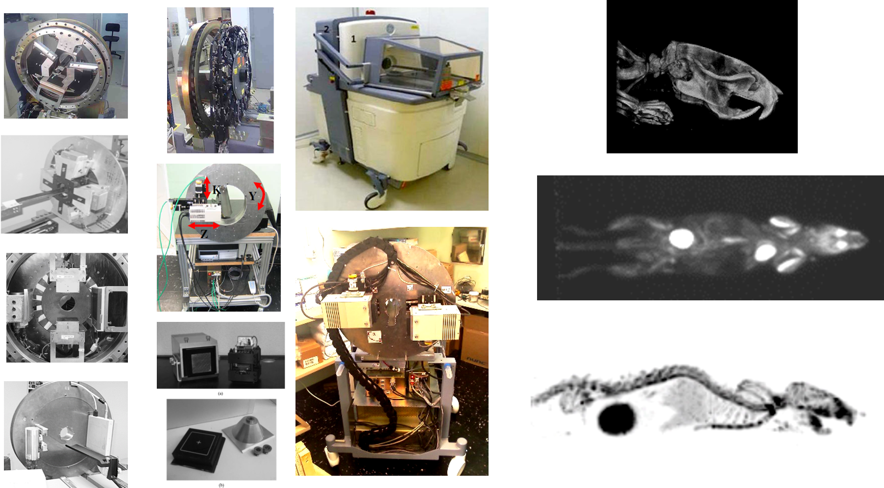
Small animal imaging is an important tool in several disciplines such as drug development and translational cancer research. The most significant among the clear advantages of imaging is that functional information at the molecular and cellular levels can be measured in an intact, living system. During his stage in the Medical Imaging Lab of the Gregorio Maranon Hopsital (IISGM) Dr Lage developed several of these systems including complete PET, PECT/CT and SPECT systems for rodent imaging.
Related links
- M. Abella, E. Vicente, A. Rodríguez-Ruano, S. Espana , E. Lage, M Desco, J.M. Udias and J.J. Vaquero. “Misalignments calibration in small-animal PET scanners based on rotating planar detectors and parallel-beam geometry”. Physics in Medicine and Biology, 57(22):7493-518, 2012, DOI:10.1088/0031-9155/57/22/7493
- Goertzen AL, Bao Q, Bergeron M, Blankemeyer E, Blinder S, Cañadas M, Chatziioannou AF, Dinelle K, Elhami E, Jans HS, Lage E, Lecomte R, Sossi V, Surti S, Tai YC, Vaquero JJ, Vicente E, Williams DA, Laforest R. “NEMA NU 4-2008 Comparison of Preclinical PET Imaging Systems”. The Journal of Nuclear Medicine, 53:1–10, 2012. DOI: 2967/jnumed.111.099382.
- M Cañadas, M Embid, E Lage, M Desco, JJ Vaquero, JM Pérez. "NEMA NU 4-2008 performance measurements of two commercial small-animal PET scanners: ClearPET and rPET(1)". IEEE Transactions on Nuclear Science, 58(1): 58-65, 2011. DOI: 1109/TNS.2010.2072935
- E Lage, J L Villena, G Tapias et al. “A SPECT Scanner for Rodent Imaging Based on Small-Area Gamma Cameras”. IEEE Transactions on Nuclear Science, 57(5): 2524-2531, 2010. DOI: 1109/TNS.2010.2057516 5-Y IF: 1.384
- M Abella, JJ Vaquero, ML Soto-Montenegro, E Lage, M Desco. "Sinogram bow-tie filtering in FBP PET reconstruction". Medical Physics, 36(5): 1663-1671, 2009. DOI: http://dx.doi.org/10.1118/1.3096707 5-Y IF: 2.954
- E Lage, JJ Vaquero, A Sisniega, et al. "Design and performance evaluation of a coplanar multimodality scanner for rodent imaging". Physics in Medicine and Biology, 54(18): 5427-5441, 2009. DOI: 1088/0031-9155/54/18/005 5-Y IF: 2.973
- JJ Vaquero, S Redondo, E Lage et al. "Assessment of a New High-Performance Small-Animal X-Ray Tomograph". IEEE Transactions on Nuclear Science, 55(3): 898-905, 2008. DOI:1109/TNS.2008.922814 5-Y IF: 1.384
- M Abella, J Vaquero, E Vicente, J Álvarez, E Lage, M Desco. "Effect of misalignments in small animal positron emission tomography scanners based on rotating planar detectors". AMI conference 2006. Poster presentation DOI: 10.1007/s11307-006-0031-x . Molecular Imaging and Biology, 2006, vol. 8, n. 2, p. 75-76, 5-Y IF: 2.76
- J J Vaquero, E Lage, S Redondo, M Abella, E Vicente, M Desco. "Initial results of a positron emission tomography/computed tomography small-animal imaging device with co-planar geometry". AMI conference 2006. Poster presentation DOI: 10.1007/s11307-006-0031-x. Molecular Imaging and Biology, 2006, vol. 8, n. 2, p. 107, 5-Y IF: 2.76
- J Pascau, J Vaquero, M Abella, R Cacho, E Lage, M Desco. "Multimodality workstation for small animal image visualization and analysis". AMI conference 2006. Poster presentation DOI: 10.1007/s11307-006-0031-x. Molecular Imaging and Biology, 2006, vol. 8, n. 2, p. 97-98, 5-Y IF: 2.76
Medical Devices
Affordable wavefront refraction technology

There is a critical need for tools that increase the accessibility of eye care to address the most common cause of vision impairment: uncorrected refractive errors. Due to the global shortage of eye care professionals, it is often difficult, particularly in low-resource settings, to obtain accurate prescriptions for eyeglasses that would effectively correct refractive errors and restore good vision. At MEDIC we have developed together with PlenOptika Inc. (MA, USA) and Aurolab (India) an innovative device that addresses this massive problem by significantly simplifying and speeding up the refraction process for prescribing eyeglasses.
- N J. Durr, S. R. Dave, F. A. Vera-Diaz, D. Lim, C. Dorronsoro, S. Marcos, F. Thorn, E. Lage. “Clinical Evaluation of a portable handheld wavefront Autorefractor”. Optometry and Vision Science (OVS) 2015, 92(12) 1140-7.
- N. Durr, S. R. Dave, E. Lage, S. Marcos, F. Thorn and D. Lim. “From unseen to seen: tackling the global burden of uncorrected refractive errors”. Annual Reviews of Biomedical Engineering, Vol. 16: 131-153
- Durr NJ, Dave SR, Lim Daryl, Sughanti M, Bama S, Mahadevan R, Ravilla S, Sanil J, Ravilla T, and E. Lage. Clinical Evaluation of a novel wavefront autorefractor on 708 Patients in a Hospital and vision Center in rural India Investigative Ophthalmology & Visual Science 2017;
- E. Lage, E. Garcia, S. Dave, M. Ramirez, L.M. Garcia, N. Alejandre-Alba, D. Lim, C. Dorronsoro, S. Marcos, N Durr. “Visual acuity evaluation with refractions prescribed by a novel low-cost wavefront aberrometer”. ARVO 2015 conference.. Investigative Ophthalmology & Visual Science June 2015, Vol.56, 3570.
- Nicholas J. Durr, Eduardo Lage, Shivang R. Dave, Carlos Dorronsoro, Susana Marcos, Daryl Lim. “Apparatus and Method for determining an eye prescription”, PCT/US2014/045261
- Lage, F. Vera-Diaz, S R. Dave, D. Lim, C. Dorronsoro, F. Thorn and N. Durr. “Evaluation of a low-cost wavefront aberrometer for measuring refractive errors”. ARVO 2014 conference. Investigative Ophthalmology & Visual Science April 2014, Vol.55,
- Plenoptika feature in MIT news Home page http://news.mit.edu/2018/startup-plenoptika-vision-care-developing-world-0111 , Jan 2018
Simultaneous Vision Simulation

SimVis will have a major clinical impact in the field of presbyopia corrections, as it holds potential to change the way these corrections are prescribed: This technology allows the patient for first time to experience the real world through multifocal corrections before surgery, so the patients can decide what correction will fit better their expectations before intra-ocular lens implantation. MEDIC is helping to develop this Technology in collaboration with the VioBio Lab (Instituto de Optica Daza de Valdes, CSIC, Spain) and the Spin-off Company 2EyesVision.
- C. Dorronsoro, X. Barcala, E. Gambra, V. Akondy, L. Sawides, Y Marrakchi, V. Rodriguez-Lopez, C. Benedi-Garcia, M. Vinas, E. Lage, S. Marcos. " Tunable Lenses: dynamic characterization and fine-tuned control for high speed applications", Optics Express (in Press)
- Yassine Marrackchi, “Development of electronics and firmware for a vision simulation device”, Bachelor Thesis, Director E. Lage, Biomedical Engineering, Universidad Carlos III de Madrid, 2017
- Telemadrid News, “Madrid reconoce a los mejores 7 trabajos de ciencia y tecnologia”, http://www.telemadrid.es/noticias/madrid/noticia/madrid-reconoce-los-7-mejores-trabajos-de-ciencia-y-tecnologia , March 2018

